
Utilité clinique de l’A-TCO
Résumé
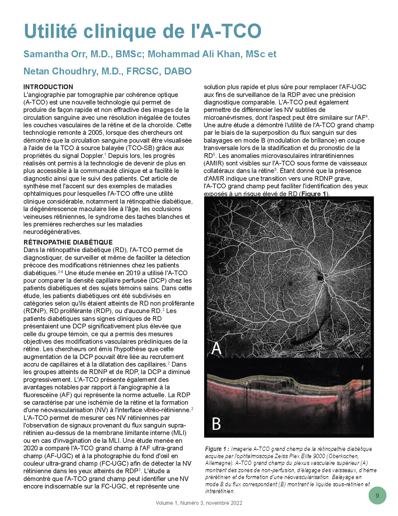
L’angiographie par tomographie par cohérence optique (A-TCO) est une nouvelle technologie qui permet de produire de façon rapide et non effractive des images de la circulation sanguine avec une résolution inégalée de toutes les couches vasculaires de la rétine et de la choroïde. Cette technologie remonte à 2005, lorsque des chercheurs ont démontré que la circulation sanguine pouvait être visualisée à l’aide de la TCO à source balayée (TCO-SB) grâce aux propriétés du signal Doppler. Depuis lors, les progrès réalisés ont permis à la technologie de devenir de plus en plus accessible à la communauté clinique et a facilité le diagnostic ainsi que le suivi des patients. Cet article de synthèse met l’accent sur des exemples de maladies ophtalmiques pour lesquelles l’A-TCO offre une utilité clinique considérable, notamment la rétinopathie diabétique, la dégénérescence maculaire liée à l’âge, les occlusions veineuses rétiniennes, le syndrome des taches blanches et les premières recherches sur les maladies neurodégénératives.
Références
Spaide RF, Fujimoto JG, Waheed NK, Sadda SR, Staurenghi G. Optical coherence tomography angiography. Prog Retin Eye Res. 2018;64:1. doi:10.1016/J.PRETEYERES.2017.11.003
Rosen RB, Andrade Romo JS, Krawitz BD, et al. Earliest Evidence of Preclinical Diabetic Retinopathy Revealed Using Optical Coherence Tomography Angiography Perfused Capillary Density. Am J Ophthalmol. 2019;203:103-115. doi:10.1016/J.AJO.2019.01.012
Pichi F, Smith SD, Abboud EB, et al. Wide-field optical coherence tomography angiography for the detection of proliferative diabetic retinopathy. Graefe’s Arch Clin Exp Ophthalmol. 2020;258:1901-1909. doi:10.1007/s00417-020-04773-x
Hwang TS, Jia Y, Gao SS, et al. Optical Coherence Tomography Angiography Features of Diabetic Retinopathy. Retina. 2015;35(11):2371-2376. doi:10.1097/IAE.0000000000000716
Arya M, Sorour O, Chaudhri J, et al. Distinguishing Intraretinal Microvascular Abnormalities from Retinal Neovascularization using Optical Coherence Tomography Angiography. Retin J Retin Vitr Dis. 2019;00(00):1-10. doi:10.1097/IAE.0000000000002671
De Oliveira Dias JR, Zhang Q, Garcia JMB, et al. Natural History of Subclinical Neovascularization in Nonexudative Age-Related Macular Degeneration Using Swept-Source OCT Angiography. Ophthalmology. 2018;125(2):255-266. doi:10.1016/j.ophtha.2017.08.030
Laiginhas R, Yang J, Rosenfeld PJ, Falcão M. Non-Exudative Macular Neovascularization – A systematic review of prevalence, natural history, and recent insights from OCT angiography. Ophthalmol Retin. 2020;4(7):651-661. doi:10.1016/J.ORET.2020.02.016
Yanagi Y, Mohla A, Lee SY, et al. Incidence of Fellow Eye Involvement in Patients With Unilateral Exudative Age-Related Macular Degeneration. JAMA Ophthalmol. 2018;136(8):905. doi:10.1001/JAMAOPHTHALMOL.2018.2154
Capuano V, Miere A, Querques L, et al. Treatment-Naive Quiescent Choroidal Neovascularization in Geographic Atrophy Secondary to Nonexudative Age-Related Macular Degeneration. Am J Ophthalmol. 2017;182:45-55. doi:10.1016/j.ajo.2017.07.009
Heiferman MJ, Fawzi AA. Progression of subclinical choroidal neovascularization in age-related macular degeneration. PLoS One. 2019;14(6). doi:10.1371/JOURNAL.PONE.0217805
Ouederni M, Khalifa MBH, Sassi H, Nefaa F, Ayed O, Cheour M. Quantitative Analysis of Microvascular Network with Optical Coherence Tomography Angiography and its Correlation with Visual Acuity in Retinal Vein Occlusion. J Curr Ophthalmol. 2021;33:453-460. doi:10.4103/JOCO.JOCO_163_21
Mejía ME, Ríos HA, Rosenstiehl S, Rodríguez FJ. Optical coherence tomography angiography as predictor of visual outcomes in retinal vein occlusion treated with antiangiogenic therapy. Eur J Ophthalmol. 2022;0(0):1-7. doi:10.1177/11206721221099487
Tsai G, Banaee T, Conti FF, Singh RP. Optical coherence tomography angiography in eyes with retinal vein occlusion. J Ophthalmic Vis Res. 2018;13(3):315-332. doi:10.4103/jovr.jovr_264_17
Pradas M, Rodriguez-Merchante MP, Estébanez N, et al. Navigating the White Dot Syndromes with Optical Coherence Tomography (OCT) and OCT Angiography (OCT-A). Ocul Immunol Inflamm. 2022:1-11. doi:10.1080/09273948.2022.2046798
Agarwal A, Invernizzi A. The Role of Optical Coherence Tomography and Optical Coherence Tomography Angiography in the Differential Diagnosis of Posterior Uveitis. Ocul Immunol Inflamm. 2022:1-8. doi:10.1080/09273948.2022.2071743
Testi I, Modugno RL, Pavesio C. Multimodal imaging supporting the pathophysiology of white dot syndromes. J Ophthalmic Inflamm Infect. 2021;11(32):1-7. doi:10.1186/S12348-021-00261-3
Pichi F, Sarraf D, Morara M, Mazumdar S, Neri P, Gupta V. Pearls and pitfalls of optical coherence tomography angiography in the multimodal evaluation of uveitis. J Ophthalmic Inflamm Infect. 2017;7(20):1-12. doi:10.1186/S12348-017-0138-Z
Pichi F, Srvivastava SK, Chexal S, et al. En face optical coherence tomography and optical coherence tomography angiography of multiple evanescent white dot syndrome: New insights into pathogenesis. Retina. 2016;36:S178-S188. doi:10.1097/IAE.0000000000001255
Fang M, Strand K, Zhang J, et al. Retinal vessel density correlates with cognitive function in older adults. Exp Gerontol. 2021;152:1-6. doi:10.1016/j.exger.2021.111433
Wang X, Zhao Q, Tao R, et al. Decreased Retinal Vascular Density in Alzheimer’s Disease (AD) and Mild Cognitive Impairment (MCI): An Optical Coherence Tomography Angiography (OCTA) Study. Front Aging Neurosci. 2021;12:1-10. doi:10.3389/fnagi.2020.572484
Abraham AG, Guo X, Arsiwala LT, et al. Cognitive decline in older adults: What can we learn from optical coherence tomography (OCT)-based retinal vascular imaging? J Am Geriatr Soc. 2021;69:2524-2535. doi:10.1111/JGS.17272
Yan Y, Wu X, Wang X, et al. The Retinal Vessel Density Can Reflect Cognitive Function in Patients with Alzheimer’s Disease: Evidence from Optical Coherence Tomography Angiography. J Alzheimer’s Dis. 2021;79:1307-1316. doi:10.3233/JAD-200971
Wang X, Wei Q, Wu X, et al. The vessel density of the superficial retinal capillary plexus as a new biomarker in cerebral small vessel disease: an optical coherence tomography angiography study. Neurol Sci. 2021;42:3615-3624. doi:10.1007/S10072-021-05038-Z
Lee MJ, Abraham AG, Swenor BK, Sharrett AR, Ramulu PY. Application of optical coherence tomography in the detection and classification of cognitive decline. J Curr Glaucoma Pract. 2018;12(1):10-18. doi:10.5005/JP-JOURNALS-10028-1238
Wisely CE, Wang D, Henao R, et al. Convolutional neural network to identify symptomatic Alzheimer’s disease using multimodal retinal imaging. Br J Ophthalmol. 2022;106(3):388-395. doi:10.1136/BJOPHTHALMOL-2020-317659
Publié
Comment citer
Numéro
Rubrique
Licence
© Canadian Eye Care Today 2022

Cette œuvre est sous licence Creative Commons Attribution - Pas d'Utilisation Commerciale - Pas de Modification 4.0 International.
